Introduction - Classification of histopathology images
This project is about developing Deep Learning Model (DNN) for histopathology images classification.
Histopathology refers to the microscopic examination of tissue in order to study the manifestations of disease. (Source - Wikipedia)
By developing such DNN model, we can automate and improve the diagnostic process.
Notebook Content:
The following notebook consist of 3 main parts:
- Part 1 - Training different DNN models and evaluating their performences for the classification task.
- Part 2 - Plotting Confusion matrix, PCA and TNSE visualization (For best performing model from part 1).
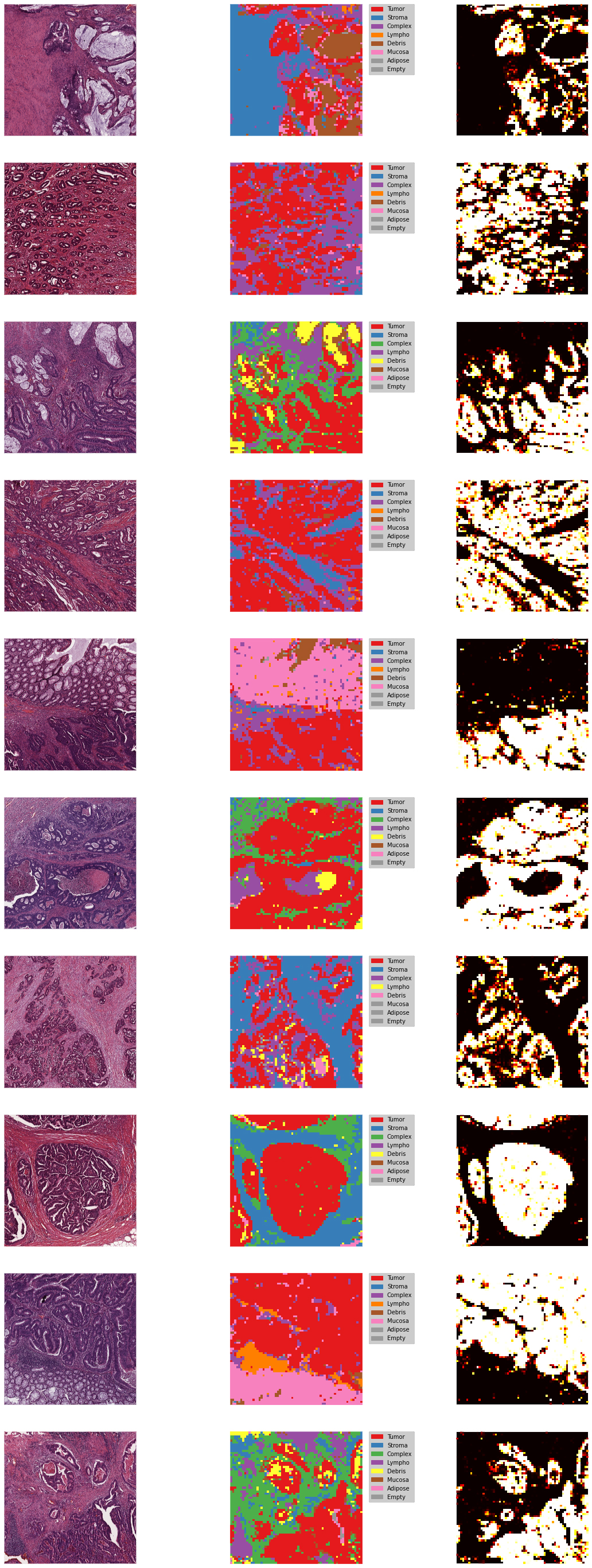
- Part 3 - Coloring Large histopathology images (5000 x 5000 x 3 RGB ), for tumor and other cells representation.
The dataset:
Colorectal histology dataset:
Classification of textures in colorectal cancer histology.
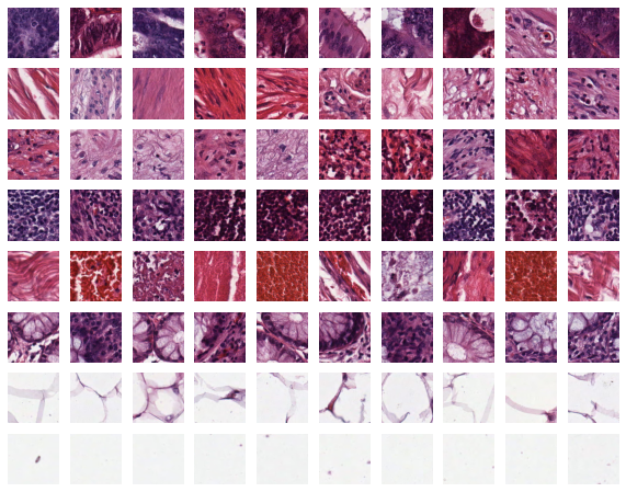
5,000 Examples. Each example is a 150 x 150 x 3 RGB image of one of 8 classes:
- Tumor
- Stroma
- Complex
- Lympho
- Debris
- Mucosa
- Adipose
- Empty
Results:
Using Transfer Learning of VGG16 model with CNN, Data Augmentation, and preprocessing:
The best model achived Accuracy score of 93.4% for cells recognition and 95.5% for Tumor recognition
Left - Original histopathology image
Middle - Classification of different cells
Right - Tumor Probability